

ABSTRACT:
Although early mobilization is essential for functional recovery after phalangeal
fractures, many fixation methods still require prolonged immobilization, increasing
the risk of joint stiffness. We herein describe a simple, safe, and low-cost technique
that combines intramedullary Kirschner wires (K-wires) with external connection, adapted
from the Ulson method, which was originally developed for distal radius fractures.
The procedure consists of closed reduction under fluoroscopy followed by the insertion
of two 1.0-mm intramedullary wires. The external ends of these wires are bent and
connected, forming a stable frame that enables immediate mobilization without the
need for immobilization. The technique provided stable fixation, reliable bone healing,
and early rehabilitation, with a low complication rate. It represents a practical
and reproducible option, especially useful in resource-limited settings.
Keywords: external fixators; finger phalanges; fracture fixation; intramedullary; fractures; bone; Kirschner wires.
RESUMO:
A mobilização precoce é fundamental para a recuperação funcional após fraturas das
falanges, mas muitos métodos de fixação ainda exigem imobilização prolongada, o que
aumenta o risco de rigidez articular. Descrevemos uma técnica simples, segura e de
baixo custo, que combina fios de Kirschner intramedulares e ligação externa, adaptada
do método de Ulson, originalmente desenvolvido para fraturas do rádio distal. O procedi-mento
consiste em redução fechada sob fluoroscopia, seguida da introdução de 2 fios intramedulares
de 1,0 mm, cujas extremidades externas são dobradas e conectadas, e formam um quadro
estável que permite mobilização imediata sem necessidade de imobilização. A técnica
proporcionou fixação estável, consolidação óssea confiável e reabilitação precoce,
com baixa taxa de complicações. Trata-se de uma opção prática e reprodutível, especialmente
útil em contextos com recursos limitados.
Palavras-chave: falanges dos dedos da mão; fios de Kirschner; fixação intramedular de fraturas; fixadores externos; fraturas ósseas.
FIGURES
| Citation: Alves GI, Severo AL, Lemos MB, Lech O, Kowaleski F, Tagliari I. Combined Intramedullary-External K-Wire Fixation for Phalangeal Fractures: Description of a Surgical Technique. 61(03):. doi:10.1055/s-0046-1824571 |
| Note: Study developed at Instituto de Ortopedia e Traumatologia (IOT), Hospital São Vicente de Paulo (HSVP), Passo Fundo, RS, Brazil. |
| Financial Support The authors declare that they did not receive financial support from agencies in the public, private, or non-profit sectors to conduct the present study. |
|
Editor-in-Chief: Geraldo da Rocha Motta Filho. |
|
Conflict of Interests The authors have no conflict of interests to declare. |
| Received: October 07 2025; Accepted: March 31 2026 |
INTRODUCTION
Phalangeal fractures of the hand are common injuries that require precise management to restore function and prevent stiffness. Stable fractures often respond well to conservative treatment, whereas unstable or displaced fractures require surgical stabilization. Traditional methods, such as Kirschner wires (K-wires), screws, and miniplates, have limitations related to prolonged immobilization, greater soft tissue dissection, or higher costs in resource-limited settings.1,2 To overcome these limitations, we herein describe a simple and accessible technique that combines intramedullary and external fixations with K-wires, providing stable alignment and enabling early mobilization in patients with fractures of the proximal and middle phalanges.3,4
SURGICAL TECHNIQUE
The technique is indicated for extra-articular diaphyseal or metaphyseal fractures of the proximal and middle phalanges that require surgical stabilization, in which minimal soft-tissue dissection and early mobilization are desired.5,6
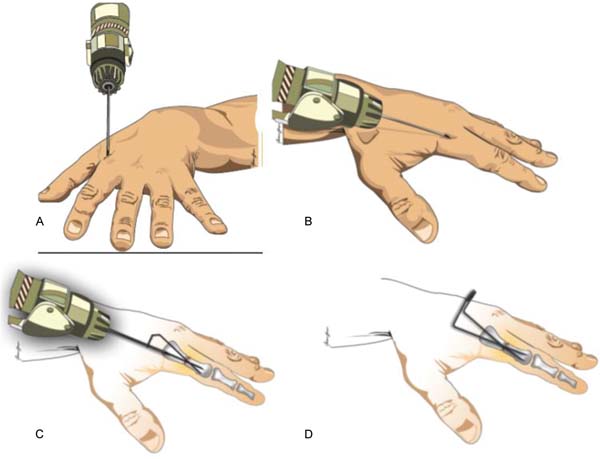
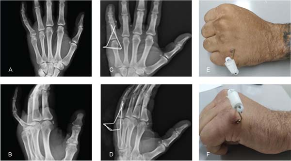
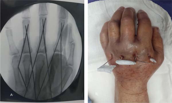
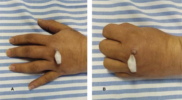
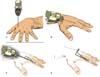
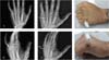
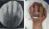
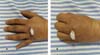
The procedure may be performed under regional or general anesthesia, with the patient in the supine position and the upper limb supported on an auxiliary table. Closed reduction is achieved under fluoroscopic guidance, applying gentle traction and digital manipulation. Two 1.0-mm K-wires are inserted intramedullarily from medial and lateral entry points at the base of the phalanx, crossing the fracture line and maintaining parallel alignment. Depending on the fracture pattern, the wires may also be inserted retrogradely. The external ends are bent to form a rectangular structure, and they are connected with microporous tape, providing additional stability. Reduction and fixation are confirmed by fluoroscopy, and only a simple dressing is applied, without the need for immobilization (►Figs. 1-4). This construct is based on the Ulson method, which was originally described for distal radius fractures and adapted here for finger frac-tures. Our technique enables K-wire insertion in a parallel configuration or crossing within the diaphysis, depending on the fracture pattern and bone size. In both configurations, the biomechanical principle remains the same, functioning as an elastic system that generates centripetal compression at the fracture site. Simple dressings are used for postoperative comfort.
Active and passive finger mobilization begin immediately after surgery. Serial radiographs are obtained every 2 weeks to monitor bone healing. The K-wires are removed after radiographic confirmation of bone healing, usually between the fifth and sixth postoperative weeks. After wire removal, the patient continues range-of-motion exercises until com-plete recovery.
OUTCOME
This technique was applied in a consecutive series of 18 patients with proximal and middle phalangeal fractures, most frequently involving the little and ring fingers. Fifteen patients completed the clinical follow-up. All fractures achieved bone healing between 5 and 6 weeks, with early recovery of mobility and a low complication rate. Three cases of superficial pin tract infection were managed conserva-tively, without loss of reduction, need for hardware removal, or delayed healing.7
Combined intramedullary-external fixation provides greater rotational stability while preserving the simplicity and minimally-invasive nature of percutaneous fixation. The external connection of the wires enables early mobilization without the need for immobilization, which helps reduce joint stiffness and lowers the risk of tendon adhesions. Moreover, the method is low-cost and easily reproducible, supporting its routine application in different care settings.8,9 A specific aspect of this configuration is the generation of centripetal forces that promote compression at the fracture site.10 In the current study, the technique was applied to extra-articular diaphyseal and metaphyseal frac-tures of the phalanges.
Despite the favorable outcomes, strict fluoroscopic con-trol and precise wire positioning are essential to avoid intra-articular penetration. Expanded clinical experience will better define the indications and long-term outcomes of this technique.
CONCLUSION
Combined internal-external fixation with K-wires represents a safe, reproducible, and low-cost option to treat unstable proximal and middle phalangeal fractures. It enables imme-diate active mobilization and reliable bone healing, with a low complication rate. The method was conceptually adapted from the Ulson technique, which was originally developed for distal radius fractures-an important Brazilian contribution to orthopedic surgery-and its successful ap-plication in phalangeal fractures reinforces the potential for adapting established techniques to new anatomical and clinical contexts.
REFERENCES