

ABSTRACT:
OBJECTIVE To evaluate the efficacy of cement augmentation in internal fixation of trochanteric
fractures in the elderly.
METHODS A comprehensive search of the MEDLINE, SpringerLink, CENTRAL, and Scopus databases
was conducted up to February 2025 for randomized clinical trials (RCTs) comparing
internal fixation with and without cement augmentation. The review adhered to the
2020 Preferred Reporting Items for Systematic Reviews and Meta-Analyses (PRISMA) statement
guidelines. Random-effects models were used to pool effect estimates. The primary
outcomes were fixation failure and tip-apex distance (TAD). The secondary outcomes
included mortality, reoperation rate, length of hospital stay, blood loss, and operative
time. The risk of bias was assessed with the revised version of the Cochrane Risk
of Bias tool (RoB 2.0; Cochrane Collaboration), and evidence certainty, with the Grading
of Recommendations, Assessment, Development, and Evaluation (GRADE) approach.
RESULTS Five RCTs comprising 509 patients were included. Cement augmentation did not significantly
reduce fixation failure (relative risk [RR] = 1.13; 95%CI: 0.20–6.43) but significantly
improved the TAD (mean difference [MD] = -2.11 mm; 95%CI: -3.13 to -1.09; p < 0.0001). There were no significant effects on mortality (RR = 1.10; 95%CI: 0.54–2.22)
or reoperation (RR = 0.12; 95%CI: 0.01–2.09). Cement augmentation shortened the hospital
stay by 2.5 days (MD = -2.59; 95%CI: -6.87 to 1.69), but it increased blood loss (MD
= 49.01; 95%CI: -61.78 to 159.8) and operative time by 11 minutes (MD = 11.8; 95%CI:
5.91–17.69).
CONCLUSIONS Cement augmentation improves fixation quality and reduces hospitalization in elderly
patients with trochanteric fractures, but it increases blood loss and operative time.
Its use should be individualized based on patient risk profiles.
Palavras-chave: bone cements; elderly; fracture fixation, internal; hip fractures.
RESUMO:
OBJETIVO Avaliar a eficácia do aumento com cimento na fixação interna de fraturas trocantéricas
em idosos.
MÉTODOS Uma busca abrangente nas bases de dados MEDLINE, SpringerLink, CENTRAL e Scopus sobre
ensaios clínicos randomizados (ECRs) que comparavam a fixação interna com e sem aumento
com cimento foi conduzida até fevereiro de 2025. A revisão seguiu as diretrizes da
declaração Preferred Reporting Items for Systematic Reviews and Meta-Analyses (PRISMA)
de 2020. Modelos de efeitos aleatórios foram utilizados para a combinação das estimativas
de efeito. Os desfechos primários foram a falha de fixação e a distância ponta-ápice
(DPA). Os desfechos secundários incluíram mortalidade, taxa de reoperação, tempo de
internação, perda sanguínea e tempo operatório. O risco de viés foi avaliado com a
versão revisada da ferramenta Cochrane Risk of Bias (RoB 2.0; Cochrane Collaboration),
e a certeza da evidência foi analisada pelo método Grading of Recommendations, Assessment,
Development, and Evaluation (GRADE).
RESULTADOS Cinco ECRs foram incluídos, num total de 509 pacientes. O aumento com cimento não
reduziu significativamente a falha de fixação(riscorelativo[RR] = 1,13; IC95% = 0,20–6,43),
mas melhorou significativamente a DPA (diferença média [DM] = 2,11 mm; IC95% = -3,13
a -1,09; p < 0,0001). Não houve efeito significativo sobre a mortalidade (RR = 1,10; IC95% =
0,54–2,22), nem sobre a taxa de reoperação (RR = 0,12; IC95% = 0,01–2,09). O aumento
com cimento reduziu o tempo de internação em 2,5 dias (DM = -2,59; IC95% = -6,87 a
1,69), mas aumentou a perda sanguínea (DM = 49,01; IC95% = -61,78 a 159,8) e o tempo
operatório em 11 minutos (DM = 11,8; IC95% = 5,91–17,69).
CONCLUSÃO O aumento com cimento melhora a qualidade da fixação e reduz o tempo de internação
em pacientes idosos com fraturas trocantéricas, mas aumenta a perda sanguínea e o
tempo operatório. Seu uso deve ser individualizado com base no perfilde riscodopaciente.
Palavras-chave: cimentos ósseos; fixação interna de fraturas; fraturas do quadril; idoso.
FIGURES
| Citation: Sugandhi A, Guna NKSD, Yapson GYA, Suryanov IPD, Putra IGNPW. Is It Necessary to Use Cement Augmentation in Internal Fixation for Trochanteric Fractures in the Elderly? A Systematic Review and Meta-Analysis. 61(3):. doi:10.1055/s-0046-1824532 |
| Note: Data Availability Data will be available upon request to the corresponding author. |
| Financial Support The authors declare that they did not receive financial support from agencies in the public, private, or non-profit sectors to conduct the present study. |
|
Conflict of Interests
The authors have no conflict of interests to declare. |
|
Editor-in-Chief:
Geraldo da Rocha Motta Filho. |
| Received: August 16 2025; Accepted: March 31 2026 |
INTRODUCTION
Trochanteric fractures are a common orthopedic challenge in the elderly, primarily due to age-related bone fragility and osteoporosis. These fractures often require internal fixation; however, conventionalfixationmethodsmay be insufficient in severely-osteoporotic bone, leading to complications such as fixation failure, implant migration, and other complications.1,2
Some internal fixation treatments for trochanteric fractures include the Gamma3 Nail (Stryker Corporation), Trochanteric Fixation Nail–Advanced (TFNA; DePuy Synthes Inc.), Proximal Femoral Nail Antirotation (PFNA; DePuy Synthes Inc.), Intertrochanteric Antegrade Nail (INTERTAN; Smith & Nephew plc), and Sliding/Dynamic Hip Screw (SHS/DHS; DePuy Synthes Inc.).3 Each type of internal fixation has its own advantages and disadvantages. Fixation failure in DHS can reach up to 8.4%,while in cephalomedullary nailing it can be as high as 6.3%.4,5 Therefore, additional techniques are needed to address this issue, one of which is the use of cement augmentation, either with polymethylmethacrylate (PMMA) or calciumphosphate cement (CPC).6,7 However, some studies suggest that cement augmentation is necessary, while others indicate that its addition is not significant and may even increase the rate of complications, such as bleeding.8–12 Based on this, further studies are needed to draw a definitive conclusion on this issue.
The present study aims to determine whether cement augmentation is necessary in internal fixation for trochanteric fractures in the elderly by evaluating its impact on fixation failure, the need for revision surgery, and other complications.
METHODS
The current meta-analysis followed the requirements of the Cochrane Handbook for Systematic Review of Interventions, and it was registered in the International Prospective Register of Systematic Reviews (PROSPERO; CRD420251006740). As the review did not constitute a clinical trial, ethical approval was not deemed necessary.
The present study included randomized clinical trials (RCT) on cement augmentation of internal fixation for trochanteric fractures in the elderly. The inclusion criteria were patients older than 65 years with trochanteric fractures, internal fixation with the TFNA, PFNA, Gamma3 nail, or SHS/DHS, and a minimum of 6 months of follow-up. And the exclusion criteria were cadaveric studies, reviews, metaanalyses, and case reports, pathologic, open, periprosthetic, or non-trochanteric fractures, and unknown follow-up, non-traumatic fractures, non-union, or revision surgeries.
Literature Search Strategy
The current study was written according to the guidelines of the 2020 Preferred Reporting Items for Systematic Review and Meta-Analyses (PRISMA) statement. Two independent clinical researchers (AS and NKSDG) searched online databases such as MEDLINE, SpringerLink, CENTRAL, and Scopus on February 24th, 2025. To minimize the risk of missing relevant research, no filters were applied to the search strategy. The detailed search strategy is provided in the Supplementary Material.
Data Extraction and Risk of Bias Assessment
Two authors (AS and NKSDG) extracted and recorded relevant data from each included study following a standardized protocol. The extracted information included the authors and year of publication, country, study period, device, type of cement, patient demographics (age, gender, fracture type), and follow-up duration. Additionally, the incidence of intraoperative complications and the need for further surgery were calculated for each study.
Two authors independently assessed the risk of bias in the included studies using the revised Cochrane Risk of Bias tool (RoB 2.0; Cochrane Collaboration). Any discrepancies were resolved through discussion, or, if necessary, consultation with a third evaluator. The RoB 2.0 evaluates five domains: bias arising from the randomization process, deviations from intended interventions, missing outcome data, measurement of outcomes, and selective reporting. Each domain is rated as presenting low risk, some concerns, or high risk of bias. An overall risk of bias assessment was determined separately for each outcome in each study, based on evaluations across all domains.
Outcome Measures
The primary outcomes were fixation failure and tip-apex distance (TAD) measurement, while the secondary outcomes included mortality, reoperation, hospital stay, blood loss, and operative time.
Data Analysis
The statistical analysis was conducted on the integrated development environment RStudio (free and open source) for the development of a meta-analysis with an evaluation of dichotomous and continuous variables. For the dichotomous outcomes—fixation failure, mortality, and reoperation—we calculated the relative risk (RR) with 95%CIs. For the continuous variables—TAD, length of hospital stay, blood loss, and operative time—we calculated the mean difference (MD) with 95%CIs.
Statistical heterogeneity was estimated using the I2 statistic and between-study variance (Tau2), with additional assessment using the Cochran’s Q test. Heterogeneity was further evaluated by visual inspection of forest plots, and the I2 statistic was interpreted as low (0–40%), moderate (30– 60%), substantial (50–90%), and considerable (75–100%). When I2 exceeded 50%, we investigated potential sources of heterogeneity. The Cochrane’s Q test was used to assess heterogeneity, with statistical significance set at p < 0.10.
We applied a random-effects model for all analyses, as it accounts for variability across studies. Additionally, we conducted sensitivity analyses to explore potential heterogeneity and assess the impact of bias on effect estimates. Statistical significance for the main analyses was set at p < 0.05.
RESULTS
Search Results
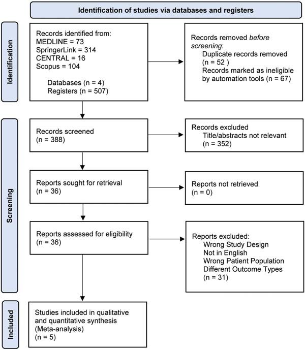
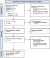
A total of 512 references were identified and assessed. A comprehensive overview of the search and selection process is illustrated in ►Fig. 1. After removal of duplicate records and records deemed ineligible by automation tools, 388 records were screened by title and abstract, 352 of which were excluded as not relevant. A total of 36 full-text articles were assessed for eligibility, and 31 were excluded due to inappropriate study design, language other than English, incorrect patient population, or different outcome measures. Ultimately, five randomized controlled trials8–12 met the inclusion criteria and were included in the qualitative and quantitative synthesis (meta-analysis).
Study Characteristics
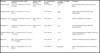
We searched 4 databases and included 5 studies8–12 (1997–2019), from Sweden, Italy, Germany, China, and Colombia, on cement augmentation in proximal femoral fracture fixation using the DHS, Gamma3, PFNA, TFNA, and the Aimstrong Proximal Femoral Nail (APFN, Double Medical Technology Inc.). As summarized in ►Table 1, PMMA was used in 3 studies,8,10,11 CPC, in one,12 and, in the remaining study,9 the material was not reported. Cement volumes ranged from 2 to 20 ml; 2 studies9,10 did not report the amounts.
| Author (year) | Country | Study period | Device | Type of cement | Cement injected | Total sample size | Gender (F/M) | Mean age (years) | A031 (Al /A2/A3)-A032A2 | Follow-up | ||||
|---|---|---|---|---|---|---|---|---|---|---|---|---|---|---|
| aug | control | aug | control | aug | control | aug | control | |||||||
| Mattsson et al.12 (2005) | Sweden | 1997-2000 | DHS | Calcium Phosphate | 20 ml | 55 | 57 | 44/11 | 47/10 | 81.2 ± 7.0 | 82.0 ±6.3 | NR | NR | 6 months |
| Dall’Oca et al.11 (2010) | Italy | January 2006-March 2010 | Gamma3 nail | PMMA | 2-3 mL | 40 | 40 | 26/14 | 30/10 | 85.3 ± 2.3 | 82.3 ±1.2 | 0/20/15 | 0/22/14 | 12 months |
| Kammerlander et al.10 (2018) | Germany | March 2012—July 2015 | PFNA | PMMA | NR | 87 | 135 | 99/19 | 86/21 | 86.1 ±4.6 | 85.6 (4.9) | 0/9/9 | 0/96/22 | 12 months |
| Ni et al.9 (2022) | China | January 2017-January 2019 | PFNA and APFN | NR | NR | 22 | 20 | NR | NR | NR | NR | NR | NR | 12-16 months |
| Salazar et al.8 (2023) | Colombia | September 2015-December 2017 | TFNA | PMMA | 4 mL | 23 | 30 | 19/4 | 23/7 | 83.87 | 85.91 | 5/15/2/1 | 8/7/12/3 | 12 months |

The augmented groups had 22 to 87 patients; the control groups had 20 to 135 subjects. Most participants were elderly female individuals (mean age: 81.2–86.1 years in the augmented groups, and 82.0–85.9 among the controls). Three studies8–10 reported the Arbeitsgemeinschaft für Osteosynthesefragen (AO) classifications (mainly AO31-A2/ A3). Follow-up generally lasted 12 months; in 1 study,12 it lasted 6 months, and, in another,9 12 to 16 months.
Risk of Bias Assessment
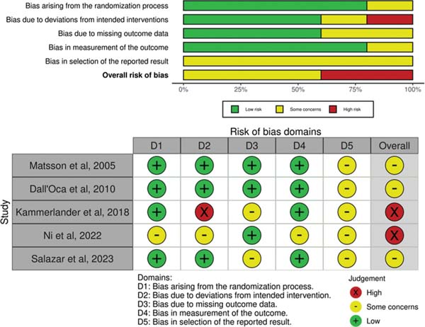
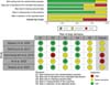
Two authors (AS and IPDS) independently assessed the risk of bias for the outcomes of fixation failure and TAD using the RoB 2.0 tool (►Fig. 2). Three studies presented low risk of bias or some concerns, while two9,10 were deemed to have a high overall risk of bias, mainly related to deviations from intended interventions and outcome measurement. Bias arising from the randomization process and missing outcome data was generally low. As commonly observed in surgical trials, blinding-related domains represented the main sources of potential bias; however, the primary outcomes, fixation failure and TAD, are objective measures and are, therefore, less susceptible to performance and detection bias. The presence of high-risk studies and small sample sizes may have contributed to imprecision in several outcomes, and the overall certainty of evidence should be interpreted with caution.
Meta-analysis Results
The comparison of the outcomes of cement augmentation in internal fixation for trochanteric fractures are summarized in ►Table 2 and in ►Table S3 of the Supplementary Material.
| Outcome | Anticipated absolute effects (95%CI) | Relative effect (95%CI) | No. of participants (studies) | Certainty of evidence (GRADE) | Comments |
|---|---|---|---|---|---|
| Fixation failure | 2 more per 1,000 (from 14 fewer to 98 more) | RR 1.13 (0.20-6.43) | 387 (3 RCTs) | Lowa,b | Very uncertain about the effect on fixation failure |
| Tip-apex distance | MD = 2.11 mm lower (3.13-1.09 lower) | - | 245 (3 RCTs) | Higha | Likely reduction in TAD with cement augmentation |
| Mortality (1 year) | 9 more per 1,000 (from 42 fewer to 112 more) | RR 1.10 (0.54-2.22) | 302 (2 RCTs) | Lowa,b | Little to no difference in mortality |
| Reoperation | 39 fewer per 1,000 (from 44 fewer to 48 fewer) | RR 0.12 (0.01-2.09) | 222 (2 RCTs) | Lowa | Cement augmentation may reduce the reoperation rate |
| Length of hospital stay | MD = 2.59 days lower (6.87 lower to 1.69 lower) | - | 234 (3 RCTs) | Lowa,c | Cement augmentation may slightly reduce the length of hospital stay |
| Blood loss | MD = 49.01 ml higher (61.78 lower to 159.8 higher) | - | 154 (3 RCTs) | Lowa,c | Evidence uncertain for blood loss difference |
| Operative time | MD = 11.8 min higher (5.91-17.69 higher) | - | 234 (3 RCTs) | Moderatea,c | Cement augmentation increases the operative time |
Primary Outcomes
Fixation Failure
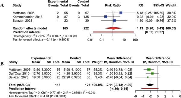
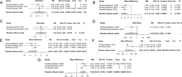
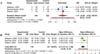
Data from 3 studies8,10,12 involving 387 participants were combined in this meta-analysis to assess fixation failures. In the cement augmentation group, one case of cut-through was reported,8 along with two cases of plate loosening.12 The pooled analysis showed no significant benefit of cement augmentation, with a wide confidence interval (RR = 1.13; 95%CI: 0.20–6.43; p = 0.8903) The certainty of evidence was rated as low. Additionally, there was no indication of substantial statistical heterogeneity (Tau2 = 0.1897; I2 = 7.6%) (►Fig. 3A; ►Supplementary Material, Fig. S1).
Tip Apex Distance
Three studies8,11,12 (245 participants) reported data on TAD. The analysis showed that the cement augmentation group had a significantly lower TAD compared to the non-augmented group, with a mean difference of -2.11 mm (95%CI: -3.13 to -1.09; p < 0.0001). This indicates that cement augmentation may improve the accuracy of implant placement. There was no heterogeneity among the included studies (Tau2 = 0; I2 = 0%; p = 0.6796) (►Fig. 3B; ►Supplementary Material, Fig. S2).
Secondary Outcomes
Mortality
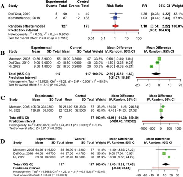
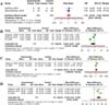
Two studies10,11 provided data on one-year mortality, which were combined for ease of analysis. The mortality rate in the cement augmentation group was 10.2%, compared to 9.1% in the non-augmented group. However, there was no statistically significant difference in postoperative mortality between the two groups (RR = 1.10; 95%CI: 0.54–2.22; p = 0.7916; I2 = 0%) (►Fig. 4A).
Reoperation
Reoperation was reported in two studies.10,12 However, only one study10 provided this outcome separately for both treatment groups, showing no significant difference between them (RR = 0.12; 95%CI: 0.01–2.09; p = 0.1454) (►Fig. 4B) and the evidence was considered of low quality. Heterogeneity was not applicable in this case.
Length of Hospital Stay
Data from 3 studies9,11,12 (117 participants in each group) were included in the meta-analysis to compare the length of hospital stay between the cement augmentation group and the control group. The results showed an MD of -2.59 days in favor of the cement augmentation group; however, this difference was not statistically significant (MD = -2.59, 95% CI: -6.87 to 1.69; p = 0.2358). The prediction interval ranged widely, from -21.07 to 15.89, indicating considerable vari-ability in the possible true effect across different settings. Substantial statistical heterogeneity was observed (Tau2 = 13.6720; I2 = 95.9%; Chi2 = 48.28; degrees of freedom [df] = 2; p < 0.0001) (►Fig. 4B).
Blood Loss
Two studies9,12 (77 participants per group) reported intra-operative blood loss. The pooled results showed an MD of 49.01 mL in favor of the control group; however, this difference was not statistically significant (MD = 49.01, 95%CI: -61.78 to 159.80; p = 0.3859). The prediction interval was wide (-1094.00 to 1192.02), indicating a high degree of uncertainty regarding the true effect across different settings. Moderate to substantial heterogeneity was observed among the included studies (Tau2 = 4896.8879; I2 = 70.8%; Chi2 = 3.43, df = 1; p = 0.0642) (►Fig. 4C).
Operative Time
A total of 3 studies9,11,12 (117 participants per group) time.9,11,12 reported on operative The pooled analysis showed a statistically significant longer operative time in the cement augmentation group compared to the non-augmented group, with an MD of 11.80 minutes (95% CI: 5.91– 17.69; p < 0.0001). The prediction interval ranged from -9.23 to 32.84, suggesting some variability in the true effect across settings. Moderate heterogeneity was detected (Tau2 = 14.8685; I2 = 53.0%), though it was not statistically significant (Chi2 = 4.25; df = 2; p = 0.1192) (►Fig. 4D).
Publication Bias
Due to the small number of included studies8–12 (n = 5), a formal assessment of publication bias using funnel-plot visualization or statistical tests such as the Egger’sor Begg’s tests was not performed. According to Cochrane Handbook recommendations, at least 10 studies are generally required to adequately detect asymmetry and assess publication bias. With fewer studies, funnel plots and regression-based methods lack sufficient power and can lead to misleading interpretations. Therefore, since the publication bias cannot be formally evaluated in the current analysis, it is a potential limitation of the present findings.
Sensitivity Analysis
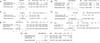
A sensitivity analysis using the leave-one-out approach was performed to evaluate the robustness of the pooled estimates across all outcomes (►Fig. 5). The analysis demonstrated that the significant reduction in TAD (MD = -2.11; 95%CI: -3.13 to -1.09; p < 0.0001) and the increase in operative time (MD = 11.80; 95%CI: 5.91–17.69; p < 0.0001) remained stable, indicating consistent findings regardless of which study was omitted. In contrast, outcomes such as fixation failure (RR = 1.13; 95%CI: 0.20–6.43; p = 0.89), mortality (RR = 1.10; 95%CI: 0.54–2.22; p = 0.79), reoperation (RR = 0.12; 95%CI: 0.01–2.09; p = 0.15), length of hospital stay (MD = -2.59; 95% -6.87 to 1.69; p = 0.24), and blood loss (MD = 49.01; 95% -61.78 to 159.80; p = 0.39) showed no statistically significant effects and varied in heterogeneity, with length of hospital stay and blood loss exhibiting particularly high between-study variance (I2 = 95.9% and 70.8% respectively). These findings suggest that, while certain operative parameters such as TAD and operative time are reliably affected by the intervention, other outcomes may be influenced by study-specific factors or lack sufficient power to detect a meaningful difference.
DISCUSSION
The current meta-analysis found that cement augmentation in internal fixation for trochanteric fractures significantly improved implant positioning, as indicated by reduced TAD. However, there was no statistically significant benefitin reducing fixation failure, reoperation, or mortality. As a result, while the mechanical stabilization hypothesis is partially supported, the overall hypothesis that cement augmentation improves clinical outcomes is only partially accepted.
The statistically significant improvement in TAD reflects a mechanical benefit that may support better implant anchorage. However, this did not translate into clinically significant differences in key outcomes such as reoperation rates or length of hospital stay, which showed no significant change. The difference in operative time was clinically modest but statistically significant. Thus, the mechanical benefitof augmentation may be clinically relevant only in select high-risk cases with unstable fractures or poor bone quality.
Compared to previous observational and biomechanical studies which showed more definitive benefits of cement augmentation in preventing implant migration and cut-out, our analysis of RCTs yielded more cautious conclusions. Studies such as those by Kammerlander et al.10 and Salazar et al.8 suggested improved construct stability, but their findings did not consistently demonstrate outcome improvements when subjected to rigorous meta-analysis. This highlights a gap between theoretical/mechanical advantages and measurable clinical effectiveness.
The strength of the current review lies in its adherence to the PRISMA guidelines, robust methodology, and sensitivity analysis. However, the conclusions are limited by the small number of high-quality RCTs and heterogeneity across studies. Future research should focus on stratifying patients based on fracture stability and bone quality, using standardized outcome measures such as functional mobility scores and exploring cost-effectiveness to guide targeted clinical application.
CONCLUSION
Cement augmentation in internal fixation for trochanteric fractures significantly reduced the TAD, supporting improved implant positioning. However, it did not significantly reduce fixation failure, reoperation rates, or one-year mortality. Similarly, no statistically significant differences were found in terms of length of hospital stay, intraoperative blood loss, or the overall reoperation rate. Operative time was significantly longer in the augmentation group. Based on these findings, the hypothesis that cement augmentation improves clinical outcomes beyond mechanical positioning is not supported.
REFERENCES